
Immunomodulatory Effect of HER2/ERBB2 Peptide on THP-1 Derived Macrophages
Publication Metrics:

Berkin Kaya1, Ayşe Nalbantsoy2*
1Department of Biotechnology, Graduate School of Natural and Applied Sciences, Ege University, Izmir, Türkiye
2Department of Bioengineering, Faculty of Engineering, Ege University, Izmir, Türkiye
*Corresponding: Ayşe Nalbantsoy, ayse.nalbantsoy@ege.edu.tr, analbantsoy@gmail.com
¹ ORCID: https://orcid.org/0009-0004-5149-2049
² ORCID: https://orcid.org/0000-0002-3852-1974
Received: 03rd October 2026
Accepted: 14th October 2026
Published: 21st October 2026
Cite This Article:
© 2026 The Author(s). This is an open-access article distributed under the terms of the Creative Commons Attribution 4.0 International License (CC BY 4.0), which permits unrestricted use, distribution, and reproduction in any medium, provided the original work is properly cited.
Abstract
Human epidermal growth factor receptor 2 (HER2) is overexpressed in approximately 30% of breast cancers and associated with aggressive behavior and poor prognosis. In addition to its established role in tumor growth and metastasis, HER2 may also influence the tumor microenvironment by modulating immune cells. Macrophages play an important role in tumor development and suppression depending on their phenotype. Due to the critical role of macrophages in cancer progression, understanding how HER2 directly affects macrophage polarization is particularly important. Therefore, this study aims to investigate the direct effect of HER2 on macrophage polarization by assessing its surface marker regulation. THP-1 human monocytic cells were differentiated into macrophages by incubation with phorbol 12-myristate 13-acetate (PMA). The cells, then were treated with different concentrations of HER2. Following treatment, M1 and M2 macrophage markers were analyzed using flow cytometry to determine the effect of HER2 on macrophage polarization. HER2 was shown to promote M1 macrophage polarization, which has pro-inflammatory and anti-tumoral features. This immunomodulator effect of HER2 indicates the its ability of immune modulation, suggesting potential use of HER2 as an adjuvant and providing a basis for future studies.
Keywords: Adjuvant, HER2, Immunomodulator, Monocyte-derived macrophages
Introduction
The ErbB family of receptor tyrosine kinases comprises four members, HER1, HER2, HER3 and HER4, which regulate cellular processes such as proliferation, migration and survival. HER2 (human epidermal growth factor receptor 2), a member of the tyrosine kinase receptor family, may promote the tumor progression by signaling between cancer cells as a network receptor. In addition, HER2 dimerizes with other RTK members, HER1, HER3 and HER4, through activation of the PI3K/Akt and Ras/Raf/MEK/MAPK pathways, which results in cell growth and differentiation.[1-4]
HER2 is overexpressed in approximately 30% of breast cancers and associated with poor prognosis; drug resistance, high metastasis rates, mediating signal transduction and allowing tumor proliferation. According to previous studies, it is also been reported that, compared to other subtypes, HER2-overexpressed breast cancer cells are expressing greater number of PD-L1 (program death ligand 1) and TILs (tumor infiltrating lymphocytes), which is involved in immune escape.[1,4,5] Given the prognostic and therapeutic decision-making, its screening has been made mandatory by the 2007 ASCO (American Society of Clinical Oncology) guideline.[6] Furthermore, accurate determination of HER2 expression has become a regular part of diagnostic evaluation of breast cancer.
In recent years, immunotherapy and HER2-targeted treatment methods have been focused on to overcome poor prognosis.[5,7] Herceptin (trastuzumab) is one of the HER2-targeted therapies, which is primarily used for HER2+ breast cancer; it inhibits the HER2 signaling pathways, which are crucial for tumor progression.[8] Trastuzumab initiates immune response, increase antibody-dependent cellular phagocytosis (ADCP) by macrophages and enhance the activity of NK cells against HER2+ tumor cells [9,10]. However, in clinical practice, a significant quantity of patients develop resistance to HER2-targeted therapies, which is thought to be mostly attributed to the phosphorylation of the HER2 receptor. The resistance may be considered multifactorial and may involve not only tumor cells but also the immune cells surrounding them.
Consequently, researchers have shifted their focus towards not only improving HER2-targeted therapies but also enhancing the immune response. Moreover, previous studies have mainly focused on the activation of antigen-presenting DCs (dendritic cells), which are stimulated by HER2 and play a critical role in initiating and modulating the antitumor immune response.[11,12] However, how HER2 interacts with other cells of the innate immune system, particularly macrophages, is of growing interest.
HER2 has an important role in the tumor microenvironment; regulating immune cells including macrophages, which may promote tumor development depending on macrophage phenotype.[9,13] Macrophages, the effector cells of the immune system, are derived from monocyte differentiation of myeloid cells, which originate from hematopoietic stem cells in the bone marrow have significant roles in immune response against cancer.[14,15] Macrophages are polarized by undergoing phenotypic changes to M1 or M2 depending on the stimuli.[16] M1 macrophages have pro-inflammatory features, initiate inflammation and antigen presentation, play role in the activation of T and natural killer (NK) cells, generate the immune response and suppress cancer development, in contrast M2 macrophages have anti-inflammatory features and play role in wound healing, angiogenesis, immune suppression and metastasis.[16-18] Tumor-associated macrophages (TAMs) frequently resemble the M2 phenotype, thereby supporting tumor growth. The polarization state can be characterized by expression of surface markers, such as CD80 and CD86 for M1 macrophages and CD163 and CD206 for M2 macrophages.[16-18] Therefore, influences on macrophage polarization may play a role in shaping the fate of cancer cells regarding its phenotype.
Although HER2-driven immune responses have been widely investigated, to date its impact on macrophage polarization still remains unclear. The THP-1 cell line is widely used as an in vitro model for macrophages, since it can be differentiated into macrophages and subsequently polarized toward M1 or M2 phenotypes. Therefore, this in vitro study addresses this gap by investigating the effect of HER2 on THP-1 derived macrophage modulation through evaluating its surface marker induction, highlighting new insight into its immunomodulatory potential and future studies.
Materials and Methods
Cell culture
THP-1 (human monocytic cell) was obtained from ATCC (TIB-202, Manassas, VA, USA). The cell line was checked before use to be mycoplasma-free. The cells were cultivated in RPMI1640, supplemented with10% fetal bovine serum (FBS, heat-inactivated twice), 2 mM L-glutamine, 100 µg/mL streptomycin and 100 U/mL penicillin (Gibco, New York, NY, USA) and incubated in a humidified atmosphere at 37°C, 5% CO2.
Differentiation of THP-1 cells into m0 macrophages
The HER2 (HER2/ERBB2 MW 70.6 kDa, Catalog No: ER2-M5220, Acro Biosystems, Newark, DE) effect was examined on phorbol 12-myristate 13-acetate (PMA, Sigma, St. Lois, MO, USA) induced THP-1-derived M0 macrophages. For this purpose, THP-1 cells were cultivated in 6-well plate a concentration of 1 × 10⁶ cells/mL in 1 mL medium with 15 ng/mL PMA with medium and incubated at 5% CO2, 37 °C for 48 h. The control group was treated only with medium.[19] Then, differentiation of M0 macrophage was evaluated by assessing microscopy and CD11b expression (Figure 1 and Figure 2).
HER2 effect on macrophage polarization
The M0 macrophages were treated with HER2 (5 and 10 µg/mL) for 24 h—which is a timeframe frequently used in macrophage polarization studies—and then cells were collected by trypsinization.[19-21] Following, cells were washed with cold Phosphate Buffered Saline (PBS, Gibco, New York, NY, USA) for 2 times. Then, 1 µL of FITC-conjugated anti-human CD11b, CD163, and PE-conjugated anti-human CD80 (Biolegend, San Diego, CA, USA) was added to cell for staining, and incubated for 45 min at room temperature in the dark. Following, cells were washed with 1 mL of PBS and analyzed immediately by using flow cytometry (BD Accuri, C5, Franklin Lakes, NJ, USA). All experiments were conducted with three independent replicates, each in triplicate.
Statistical analysis
GraphPad Prism 8.4.2 software (GraphPad 8.4.2, La Jolla, CA, USA) was used for statistical analysis. One-way ANOVA analysis was performed and followed by Dunnet posttest for comparation of results and control groups, p values of <0.05 were considered as statistically significant.
Results
Determination of THP-1 induced M0 macrophages
The immunomodulatory effect of HER2 on macrophage polarization was determined through flow cytometry analysis, based on the surface receptor expression quantified by Mean Fluorescence Intensity (MFI) values. Therefore, THP-1 cells were used to generate M0 macrophages via incubation with 15 ng/mL PMA for 48 hours and observed under a microscope (Figure 1). Following the PMA treatment, CD11b surface marker expression was determined 78.7%, which showed the successful differentiation of THP-1 cells into M0 macrophages (Figure 2).
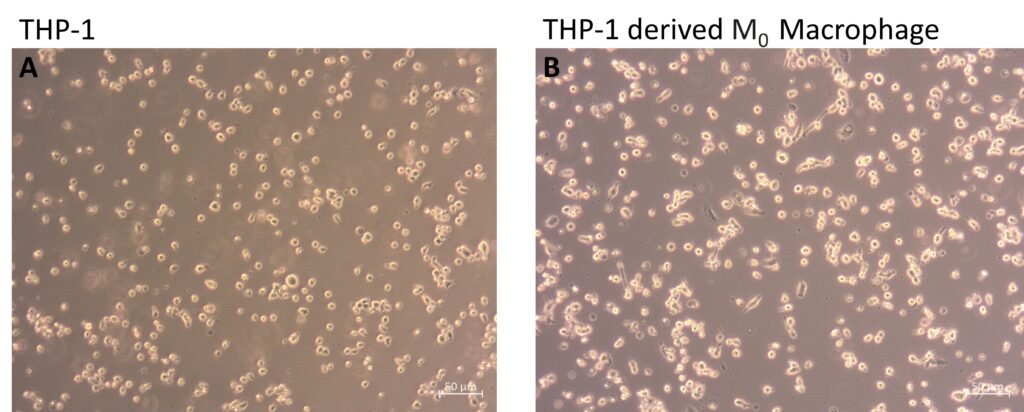
Figure 1. Microscopic images of differentiation of THP-1 cells to M0 macrophage by incubation with 15 ng/mL PMA (phorbol 12-myristate 13-acetate). (A) THP-1 cells, (B) THP-1 derived M0 macrophages.
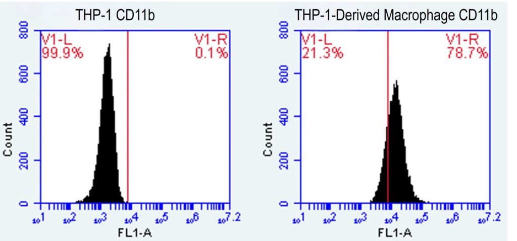
Figure 2. Differentiation of THP-1 cells to M0 macrophage by incubation with 15 ng/mL PMA (phorbol 12-myristate 13-acetate) were evaluated with CD11b (M0 macrophage) surface marker expression.
Effect of HER2 on macrophage surface markers
The mean fluorescence intensity (MFI) values of macrophages, either untreated or treated with HER2, were analyzed by flow cytometry to evaluate macrophage polarization. While CD163 expression does not increase, which indicates that HER2 did not promote M2 macrophages, in contrast, CD80 expression, which is the marker of M1 macrophages, was significantly increased after treatment, demonstrating that HER2 promoted to M1 macrophage polarization (Figure 3).
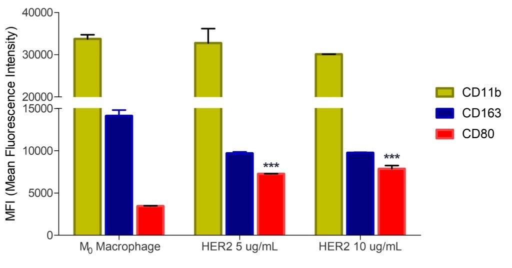
Figure 3. HER2 effect on macrophage polarization was analyzed through surface markers CD11b (M0 macrophage), CD80 (M1 macrophage) and CD163 (M2 macrophage) by flow cytometry and demonstrated with MFI (mean fluorescence intensity) ± SD.
As shown in Table 1, both HER2 concentrations of 5 and 10 µg/mL increased CD80 expression to 7272.64 ± 10.42 and 7867.20 ± 346.9, respectively. Both 5 µg/mL and 10 µg/mL HER2 treatments significantly increased CD80 expression compared to the control group (M0 macrophage) (p < 0.001***), whereas no statistically significant differences between 5 and 10 µg/mL HER2 concentrations were observed.
It is also observed that, while CD80 expression significantly increased after HER2 treatment, CD163 expression decreased: MFI values at 5 and 10 µg/mL were 9691.38 ± 189.24 and 9761.85 ± 47.15 (p < 0.001***), respectively, compared to control (14112.40 ± 672.7) (Table 1).
| Table 1. MFI values of THP-1 derived M0 macrophage and treated with HER2. The results are compared to the control group (M0 macrophage), and data are presented as mean ± SD (p <0.001***). | |||
| CD11b | CD163 | CD80 | |
| M0 Macrophage | 33173.90 ± 1636.64 | 14112.40 ± 672.7 | 3451.64 ± 36.29 |
| HER2 5 µg/mL | 32765.10 ± 1923.2 | 9691.38 ± 189.24*** | 7272.64 ± 10.42*** |
| HER2 10 µg/mL | 30114.70 ± 13.23 | 9761.85 ± 47.15*** | 7867.20 ± 346.9*** |
| MFI = Mean Fluorescence Intensity | |||
In addition, although CD80 and CD163 expression significantly changed after HER2 treatment, CD11b only decreased slightly, not statistically significant. Compared to M0 macrophage MFI values (33173. ± 1636.64), the determined MFI values of HER2 treatment at 5 and 10 µg/mL were 32765.10 ± 1923.2 and 30114.70 ± 13.23, respectively (Table 1).
Discussion
In this study, the potential use of HER2 as an immunomodulator was investigated by its direct effect on macrophage polarization under in vitro conditions. Approximately 30% of breast cancer cases overexpress HER2, which belongs to the tyrosine kinase receptor family and promotes tumor growth by acting as a signaling receptor between cancer cells.[1,4,5]
The HER2 effect on macrophage polarization was determined via its surface markers, which were finalized by flow cytometry analysis. Furthermore, for this purpose, THP-1 cells were used, since THP-1 cells can be differentiated into M0 macrophages by exposure to PMA and it is a well-established and frequently used model for studying macrophage phenotype and functions.[19,20]
After exposure of PMA, CD11b expression increased drastically, which is shows differentiation into M0 macrophages (Figure 2).
In the study conducted by Özverel et al.(2020), bone marrow-derived dendritic cells were activated with HER2 and tumor progression was prevented by inducing an immune response against the tumor.[11] A similar result was demonstrated in dendritic cells loaded with HER2-VLP (virus-like particle) fusion.[11] A monoclonal antibody Herceptin primarily used in HER2-expressed breast cancer and in the scope of macrophage, Herceptin promotes infiltration of M1 macrophage in tumor tissue. It has also been shown that Herceptin treatment promotes ADCP by activation of NK cells.[9,10,22] Overall, while the effects of HER2 on various immune cells, including dendritic cells and NK cell–mediated ADCP have been investigated in previous studies, its direct effect on macrophage polarization has yet to be elucidated.
Macrophages are the effector cells of immune system that promote or reduce tumor growth, depending on their type.[16,17] M1 macrophage has the ability to initiate immune responses in cancer treatments by initiating the pro-inflammatory process.[16,23,24]
Macrophage polarization after treatment with HER2 was analyzed through its surface marker expression (CD163 and CD80), which are most widely accepted and commonly used for identification of M1 and M2 macrophages.[19] The result of this study shows that HER2 treatment significantly induces CD80 expression (Figure 3). As is known, CD80 is a marker of M1 macrophages; the findings demonstrated that HER2 treatment promoted M0 macrophages into M1 macrophages. Additionally, when comparing the two HER2 treatment concentrations, even though 10 µg/mL HER2 induced slightly more CD80 expression, there was no significant difference in M1 macrophage polarization between 10 and 5 µg/mL treatment (Table 1). This finding suggested that lower concentrations of HER2 could be used in further studies, in any case of toxicity. Nevertheless, in addition to flow cytometry analysis, future studies should also evaluate relevant cytokines parallel to surface markers for better understanding HER2 effect on macrophage polarization.
This immunomodulatory effect of HER2 indicates that HER2 may have the potential to be used as an adjuvant due to its ability to induce pro-inflammatory M1 macrophage polarization, which has a critical role to enhance immune system and generating an anti-tumoral response. Although these findings highlight the potential of HER2 on immunomodulation, future studies should focus on evaluating its mechanism, as well as relevant pathways for better understanding.
Flow cytometry analysis demonstrated that HER2 did not promote M2 macrophage polarization, as MFI values of M2 macrophage marker CD163 decreased following to HER2 treatment (Figure 3). M2 macrophage, also known as tumor-associated macrophage (TAM), are associated with the progression of tumor growth and metastasis through mechanisms such as angiogenesis and escape from the immune system.[16,17,25] It is also known that high ratio of M2/M1 macrophages may result in poor prognosis in breast cancer, as TAMs inhibit antigen presentation features of DCs, suppress cytotoxic T cell and recruit Tregs in the tumor microenvironment.[26,27]
The fact that HER2 did not induce M2 but promoted M1 macrophage polarization is a positive sign that it may have the ability to stimulate the immune system to initiate pro-inflammatory and anti-tumoral response; therefore, HER2 may have potential use as an immunomodulatory agent and adjuvant and should be studied further for its interaction with macrophages and the mechanism involved.
Conclusion
The results of this study demonstrated that HER2 exhibits extensive immunomodulatory effects on macrophages by promoting pro-inflammatory M1 macrophage polarization, as evidenced by the upregulation of CD80 marker, without inducing anti-inflammatory M2 macrophage. These findings indicate the immunomodulatory ability of HER2 toward innate immune activation, and it suggests that it could serve as a potential adjuvant candidate and anti-tumoral agent in cancer therapy. Nevertheless, these findings provide a basis for future mechanistic and preclinical studies.
Author Contributions
Berkin Kaya: Conceptualization, methodology, analysis, investigation, writing – original and first draft.
Ayşe Nalbantsoy: Conceptualization, methodology, analysis, resources, writing – original draft, supervision.
All authors have read and approved the final version of the manuscript.
Data Availability Statement
Datasets of this study are not publicly available and can be provided upon reasonable request.
Conflict of Interest
All authors declare that they have no conflicts of interest.
Ethics Committee Approval
In the scope of this study, there is no reason to request ethics committee approval.
References
- Yarden Y. Biology of HER2 and its importance in breast cancer. Oncology. 2001;61(Suppl 2):1–13. https://doi.org/10.1159/000055396.
- Ahmed S, Sami A, Xiang J. HER2-directed therapy: Current treatment options for HER2-positive breast cancer. Breast Cancer (Tokyo, Japan). 2015;22(2):101–116. https://doi.org/10.1007/s12282-015-0587-x.
- Marchiò C, Annaratone L, Marques A, Casorzo L, Berrino E, Sapino A. Evolving concepts in HER2 evaluation in breast cancer: Heterogeneity, HER2-low carcinomas and beyond. In Seminars in Cancer Biology. 2020;Academic Press.
- Dvir K, Giordano S, Leone JP. Immunotherapy in breast cancer. International Journal of Molecular Sciences. 2024;25(14):7517. https://doi.org/10.3390/ijms25147517.
- Wu X, Yang H, Yu X, Qin JJ. Drug-resistant HER2-positive breast cancer: Molecular mechanisms and overcoming strategies. Frontiers in Pharmacology. 2022;13:1012552. https://doi.org/10.3389/fphar.2022.1012552.
- Krishnamurti U, Silverman JF. HER2 in breast cancer: A review and update. Advances in Anatomic Pathology. 2014;21(2):100–107. https://doi.org/10.1097/PAP.0000000000000015.
- Li SG, Li L. Targeted therapy in HER2 positive breast cancer (Review). Biomedical Reports. 2013;1:499–505. https://doi.org/10.3892/br.2013.95.
- Nanda R. Targeting the human epidermal growth factor receptor 2 (HER2) in the treatment of breast cancer: Recent advances and future directions. Reviews on Recent Clinical Trials. 2007;2(2):111–116. https://doi.org/10.2174/157488707780599375.
- Upton R, Banuelos A, Feng D, Biswas T, Kao K, McKenna K, Willingham S, Ho PY, Rosental B, Tal MC, Raveh T, Volkmer JP, Pegram MD, Weissman IL. Combining CD47 blockade with trastuzumab eliminates HER2-positive breast cancer cells and overcomes trastuzumab tolerance. Proceedings of the National Academy of Sciences. 2021;118(29):e2026849118. https://doi.org/10.1073/pnas.2026849118.
- Li F, Liu S. Focusing on NK cells and ADCC: A promising immunotherapy approach in targeted therapy for HER2-positive breast cancer. Frontiers in Immunology. 2022;13:1083462. https://doi.org/10.3389/fimmu.2022.1083462.
- Tegerstedt K, Franzén A, Ramqvist T, Dalianis T. Dendritic cells loaded with polyomavirus VP1/VP2Her2 virus-like particles efficiently prevent outgrowth of a Her2/neu expressing tumor. Cancer Immunology, Immunotherapy. 2007;56(9):1335–1344. https://doi.org/10.1007/s00262-007-0281-8.
- Özverel CS, Uyanikgil Y, Karaboz İ, Nalbantsoy A. Investigation of the combination of anti-PD-L1 mAb with HER2/neu-loaded dendritic cells and QS-21 saponin adjuvant: Effect against HER2 positive breast cancer in mice. Immunopharmacology and Immunotoxicology. 2020;42(4):346–357. https://doi.org/10.1080/08923973.2020.1775644.
- Triulzi T, Forte L, Regondi V, Di Modica M, Ghirelli C, Carcangiu ML, Sfondrini L, Balsari A, Tagliabue E. HER2 signaling regulates the tumor immune microenvironment and trastuzumab efficacy. Oncoimmunology. 2018;8(1):e1512942. https://doi.org/10.1080/2162402X.2018.1512942.
- Hao NB, Lü MH, Fan YH, Cao YL, Zhang ZR, Yang SM. Macrophages in tumor microenvironments and the progression of tumors. Clinical and Developmental Immunology. 2012;Article ID 948098. https://doi.org/10.1155/2012/948098.
- Tang X, Wang Z, Wang J, Cui S, Xu R, Wang Y. Functions and regulatory mechanisms of resting hematopoietic stem cells: A promising targeted therapeutic strategy. Stem Cell Research Therapy. 2023;14(1):73. https://doi.org/10.1186/s13287-023-03316-5.
- Martinez FO, Sica A, Mantovani A, Locati M. Macrophage activation and polarization. Frontiers in Bioscience. 2008;13:453–461. https://doi.org/10.2741/2692.
- Sica A, Larghi P, Mancino A, Rubino L, Porta C, Totaro MG, Rimoldi M, Biswas SK, Allavena P, Mantovani A. Macrophage polarization in tumour progression. Seminars in Cancer Biology. 2008;18(5):349–355. https://doi.org/10.1016/j.semcancer.2008.03.004.
- Wynn TA, Chawla A, Pollard JW. Macrophage biology in development, homeostasis and disease. Nature. 2013;496(7446):445–455. https://doi.org/10.1038/nature12034.
- Oztunc O, Sumer Okkali G, Zeinali S, Nalbantsoy A, Boke Sarikahya N. Four new triterpene saponins from Cephalaria speciosa and their potent cytotoxic and immunomodulatory activities. Scientific Reports. 2023;13(1):16964. https://doi.org/10.1038/s41598-023-44114-6.
- Chanput W, Mes JJ, Wichers HJ. THP-1 cell line: An in vitro cell model for immune modulation approach. International Immunopharmacology. 2014;23(1):37–45. https://doi.org/10.1016/j.intimp.2014.08.002.
- Genin M, Clement F, Fattaccioli A, Raes M, Michiels C. M1 and M2 macrophages derived from THP-1 cells differentially modulate the response of cancer cells to etoposide. BMC Cancer. 2015. https://doi.org/10.1186/s12885-015-1546-9.
- Mitri Z, Constantine T, O’Regan R. The HER2 receptor in breast cancer: Pathophysiology, clinical use, and new advances in therapy. Chemotherapy Research and Practice. 2012;743193. https://doi.org/10.1155/2012/743193.
- Wang YC, He F, Feng F, Liu XW, Dong GY, Qin HY, Hu XB, Zheng MH, Liang L, Feng L, Liang YM, Han H. Notch signaling determines the M1 versus M2 polarization of macrophages in antitumor immune responses. Cancer Research. 2010;70(12):4840–4849. https://doi.org/10.1158/0008-5472.CAN-10-0269.
- Xia Y, Rao L, Yao H, Wang Z, Ning P, Chen X. Engineering macrophages for cancer immunotherapy and drug delivery. Advanced Materials. 2020;32(40):e2002054. https://doi.org/10.1002/adma.202002054.
- Kashfi K, Kannikal J, Nath N. Macrophage reprogramming and cancer therapeutics: Role of iNOS-derived NO. Cells. 2021;10(11):3194. https://doi.org/10.3390/cells1011319.
- Jeong H, Hwang I, Kang SH, Shin HC, Kwon SY. Tumor-associated macrophages as potential prognostic biomarkers of invasive breast cancer. Journal of Breast Cancer. 2019;22(1):38–51. https://doi.org/10.4048/jbc.2019.22.e5.
- Mehta AK, Kadel S, Townsend MG, Oliwa M, Guerriero JL. Macrophage biology and mechanisms of immune suppression in breast cancer. Frontiers in Immunology. 2021;12:643771. https://doi.org/10.3389/fimmu.2021.643771.